
Hepatitis C, an innovative pilot project in Lazio under the banner of technology’s
Train medical health workers and nurses from Addiction Services, giving them the opportunity to perform an onsite ultrasound examination in the patient with varying degrees of liver disease from chronic hepatitis C virus infection, assisted by the specialist connected in real time streaming for the interpretation of the images and the compilation of the report. Verify the technical feasibility of video streaming of images with sufficient quality for adequate diagnostic interpretation. Verify the critical issues of an approach that is absolutely innovative and possibly exportable to other settings, with a view to centralizing at the SerD the entire pathway of screening, diagnosis and treatment of patients with substance use disorder potentially affected by hepatitis C virus.
A simplified approach
A simplified and entirely remote approach via Teleconsultation, implemented with an extremely practical and easy-to-use device. All under the banner of the message ’Don“Don”t move the patient but move the information about him”.
This and more was discussed at the Policlinico Umberto I in Rome, during the CME training course on the management of addicts with hepatitis C, organized by provider Letscom E3 with the nonconditional contribution of AbbVie. The course, entitled ‘ONSITE ECOGRAPHY IN THE LOW COMPLIANCE HEPATOPATHIC PATIENT – From theory to practice. The new technology for conducting a remote ultrasound examination’is part of ‘HAND – Hepatitis in Addiction Network Delivery,’ the nationwide networking project sponsored by four scientific societies (SIMIT, FeDerSerD, SIPaD and SITD) that from 2019 involves Addiction Services and HCV Treatment Centers afferent to different Italian cities.
This is the third of five modules
This is the third of five modules that are part of the course, a pilot training project that kicked off last Nov. 10 and will conclude next Dec. 15. Among the salient topics, they discussed how to recognize so-called focal lesions of the liver: cysts, angiomas or in the worst case scenario, hepatocarcinoma.
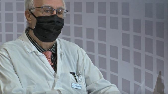
Scientific leader of the course Adriano De Santis, Associate Professor of Gastroenterology, Sapienza University of Rome, UOC Gastroenterology, Policlinico Umberto I, Rome, who addressed the motivations behind this truly cutting-edge initiative.
De Santis explained
“The initiative-explained De Santis-was born out of the need to go unearth the underground regarding chronic HCV infection. Right now therapies have allowed us to heal all of the emerged, however, we know that there is much submerged. In some categories this is very relevant.
Among these are categories with difficult management that are substance use patients who are referred to our Ser.d.”.
After clarifying that “the goal of the Course is not to teach how to interpret the image but to teach how to get the image, which will be streamed to a Center, where there will be an experienced sonographer who will interpret them, interacting and providing instructions on the best way to get an’ readable and interpretable image, De Santis then spoke about Focal Lesions, informing that “space-occupying lesions, known as LOS, are quite frequent in the general population, in most cases they are benign Los and the most common are angiomas and cysts”.
A truly cutting-edge’experience
A truly state-of-the-art experience but, as De Santis pointed out, that does not exclude the possibility that the new device may still present technical and organizational difficulties.
“We thought to make life easier for our patients to increase their compliance – said De Santis – and thus have them managed directly in the Referral Center, without having them move to the Prescribing Centers for subsequent therapies.”.
De Santis added that “in this area we have thought of training the doctors and nurses who work in the Ser.d. and we may have some difficulties with regard to interpretation, especially of ultrasound images. The availability of new equipment, therefore, of technology that allows us to perform ultrasound scans directly at the place where the patient is afferent, at low cost, only with the involvement of a specialist who can also be at a distance and, therefore, in a large hospital.”.
What this probe, the epitome of a true ‘pioneering initiative’, consists of;?
Dr. Daniela Maggi provides clarity
Providing clarity was Dr. Daniela Maggi, La Sapienza University of Rome, who together with Dr. Anna Morrone have actively contributed to the realization of the course and will play a mentoring role in the start-up phase of the project.
Maggi stated that “the mechanism is very simple. It involves a probe that connects wirelessly to the device we choose to use, which can be a tablet or a cell phone. Through this probe we are going to view the ultrasound image directly on the tablet.
Beyond the various adjustment of images and measurement of different parameters, a call can be made to an email address of the referring specialist to send the images in real time and have them viewed for interpretation at the Specialist Center.”.
Also participating in the Course was Dr. Pietro Casella, Director UOC Addictions Asl Roma 1, who highlighted the differences between patient and patient with substance use disorder.
Box disclosed
Casella disclosed that “the disease is expressed and develops in everyone in the exact same way. The elements of difference are mainly related to the elements of fragility of the patient with substance use disorder, thus difficulties inherent to the disease, which result in obstacles, often, to access to diagnosis and treatment.”.